What Does The Synaesthetic Brain Look Like? Here’s What Science Says

See colors while listening to music. Enjoy an ice cream and feel a caress on your cheek. Touching the petal of a flower and feeling a sweet taste in the mouth… All of these sensory experiences describe a neurological alteration experienced by thousands of people around the world. But what does the synaesthetic brain look like, and what actually explains this characteristic?
We know that figures such as Vincent Van Gogh, Vladimir Nabokov, Wassily Kandinsky, and Nikola Tesla were able to experience two senses at the same time. The truth is that it was long believed that synesthesia was a feature of fanciful minds and that there was no scientific basis to explain it.
However, with the arrival of the new millennium, neuroscientists, psychologists and even geneticists have become interested in this neurological singularity. What they have discovered so far is fascinating to say the least. Let’s take a closer look.
What is synesthesia?
Let’s start by clarifying the definition of synesthesia. It is a perceptual phenomenon orchestrated by a neurological disturbance. It involves the automatic and involuntary activation of several sensory or cognitive regions at the same time in response to specific stimuli. This means that the sounds can have a color, the music a flavor, the lyrics can be associated with certain tones, etc.
It is also important to take into account a number of details. Synesthesia does not respond to a hallucinatory experience, as psychiatrists Bleuler and Lehmann suspected in 1871. It also does not define a mental disorder such as schizophrenia, and is not the product of a highly imaginative mind.
It can be said that the first important step that allowed us to understand synesthesia as a neurological phenomenon was the 1995 study conducted by Dr Paulesu. In this research, different diagnostic tests were performed with positron emission tomography to measure hemodynamic responses in synaesthetes.
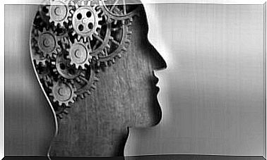
What does the synaesthetic brain look like?
It is estimated that 3 to 5% of the population suffers from some form of synesthesia. It is also more common in women than in men. On the other hand, it is interesting to know that many see it as a gift, because seeing the world through a multiple integration of several senses at the same time makes reality more intense, special and fascinating at the same time.
Not to mention another detail: it is very common for synaesthetic people to be more creative and also have better memory, because their brains have more connections. Studies such as those conducted by Dr Gian Bheeli of the University of Zurich and published in the journal Nature also indicate that there is a genetic basis and that the disease is usually inherited.
Sounds that have a sweet taste, music that bursts in color, textures that evoke images … If we wonder what the brain of a synaesthete looks like, science has already given us some interesting answers.
The phenomenon of cross-activation
As we all know, children go through a period of brain development which consists of neurological pruning. In other words, it is a process in which synaptic connections between neurons are removed in order to shape a more specialized brain.
Until the age of 12-13, it is common to have many more neurons and synapses than functionally needed. This gradual disappearance is necessary for the proper functioning of the brain.
It seems that the synaesthetes do not completely complete this neuronal pruning. Thus, different areas intersect. Research, such as that conducted at the University of Amsterdam (The Netherlands), tells us that regions associated with color in the occipito-temporal cortex can suddenly connect to motor regions, so that even some movements can evoke a tone in the person.
The limbic mediation hypothesis
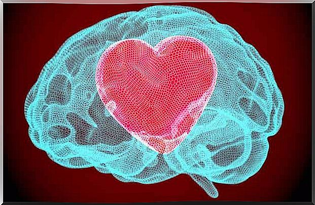
To know what the synaesthetic brain looks like, we need to consider the limbic mediation hypothesis. It was first stated by Richard Cytowic and Frank Wood in 1982 and indicates that synesthesia is orchestrated by the limbic system and, in particular, by the hippocampus.
It has been observed that synaesthetes have much more connective fibers in this area, which extend from the limbic system itself to the neocortex. This results in a greater number of perceptual phenomena, sensations, memories and even emotions.

What functional neuroimaging studies on the synaesthetic brain reveal
With the evolution of diagnostic and neuroimaging techniques, we now have much more data on what the synaesthetic brain is. So, thanks to everyone with synesthesia who volunteered for positron emission tomography (PET) and functional magnetic resonance imaging (fMRI) tests, we know the following:
- Regions of the visual cortex show increased activation.
- The density of gray matter is higher.
- There is a strong overconnectivity of the auditory cortex to the insula (the latter region is linked to emotions and the regulation of bodily homeostasis).
- As well as greater interconnectivity of the whole brain compared to non-synaesthetes.
In conclusion, synesthesia is currently a fascinating phenomenon that interests both neuroscientists and psychologists. It is expected that in the years to come we will know much more, but the most important thing is that those who present this peculiarity, far from experiencing it in a problematic way, seem to appreciate their do n.

The experiences of childhood go beyond memories or trauma, they can truly change our brains.